
Dry Eye Syndrome Stem Cell Therapy | Can Damaged Glands Actually Recover?
Waking up to eyes that feel packed with fine grit. Spending the first half hour of every morning blinking through a film that never quite clears, waiting for enough moisture to form before you can read a text message without squinting. By mid-afternoon, the light from any screen — monitor, phone, tablet — has become something to brace against rather than use.
If that description is familiar, you are not alone. Dry eye syndrome is one of the most common and persistently undertreated eye conditions in the world. According to the TFOS DEWS II international report, published in The Ocular Surface, the condition affects an estimated 344 million people globally, with prevalence climbing sharply in older adults and women after menopause. In Japan alone, credible estimates suggest over 21 million people have clinically significant dry eye, and because the condition is frequently dismissed as a nuisance rather than recognized as a chronic inflammatory disease, many patients go years without a level of treatment that matches the severity of what they actually experience.
Most people with chronic dry eye have already moved through the standard treatment sequence. Artificial tears applied throughout the day. Prescription anti-inflammatory drops — cyclosporine, lifitegrast — tried for months or years. Punctal plugs placed to prevent drainage. Heat treatment or IPL for the oil-producing glands along the eyelid margins. For mild to moderate dry eye, these approaches are genuinely useful. But for a meaningful proportion of patients — particularly those with severe aqueous-deficient dry eye, Meibomian gland dysfunction that has progressed beyond what procedural treatment can address, or dry eye driven by autoimmune conditions like Sjögren’s syndrome — the standard toolkit manages what appears on the surface while leaving the underlying gland damage untouched.
This article is written for people who have reached that point. It is for those who have been managing dry eye syndrome for years and are now asking a different question: is there a treatment that can address the cause of this, not just compensate for it?
We are a clinical team based in Malaysia with over seven years of experience in regenerative medicine, having treated more than 200 patients across a range of conditions. We will share what the peer-reviewed evidence actually shows for stem cell therapy in dry eye syndrome — including specific outcome numbers, the presentations most likely to respond, and the presentations where this treatment is unlikely to help. We will be honest about where the evidence is strong, where it is preliminary, and where stem cells simply cannot do what some clinics suggest they can.
Table of Contents
- The Gland Problem at the Heart of Dry Eye Syndrome
- Where Conventional Dry Eye Syndrome Treatments Hit Their Ceiling
- Three Biological Mechanisms Behind Stem Cell Therapy for Dry Eye Syndrome
- What Clinical Data Shows About Stem Cells and Dry Eye Syndrome
- Autologous vs. Allogeneic: Comparing Stem Cell Sources for Dry Eye Syndrome
- Who Is — and Is Not — a Good Candidate for Dry Eye Syndrome Stem Cell Therapy
- One Patient's Experience With Dry Eye Syndrome and Stem Cell Treatment
- Honest Risks and Limitations of Dry Eye Syndrome Stem Cell Therapy
- FAQ About Dry Eye Syndrome Stem Cell Treatment
- If You Have Reached the End of the Conventional Road With Dry Eye Syndrome
- References
The Gland Problem at the Heart of Dry Eye Syndrome
Why the Tear-Producing Glands in Dry Eye Syndrome Cannot Repair Themselves
The tear film that protects the surface of your eye is not simply water. It is a precisely layered structure made up of three distinct components: an outer lipid layer produced by the Meibomian glands along the eyelid margins, a middle aqueous layer produced by the lacrimal glands behind and above the eye, and an inner mucin layer produced by goblet cells on the surface of the conjunctiva. When any one of these layers is disrupted, the tear film becomes unstable — it breaks up too quickly, leaving areas of the corneal surface exposed between blinks. The result is the friction, burning, and visual fluctuation that define dry eye syndrome.
The central biological challenge is that the glands responsible for each of these layers have almost no capacity for self-repair once significant damage has occurred.
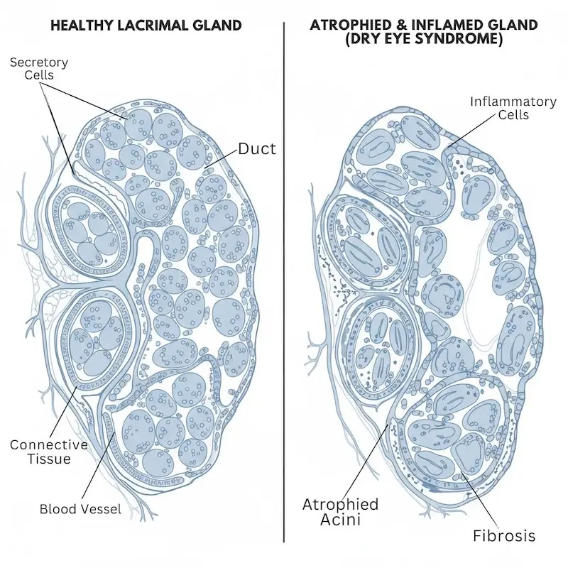
The Meibomian glands are oil-secreting structures embedded in the eyelid, approximately 25 to 30 per lid. When they become chronically inflamed or obstructed — a condition called Meibomian gland dysfunction, or MGD — the secretory cells within the gland begin to be replaced by non-functioning fibrous tissue. The glands gradually atrophy. Unlike skin, which can regenerate after injury, or the lining of the gut, which replaces itself continuously, the Meibomian glands contain only a small resident population of progenitor cells responsible for maintaining glandular tissue. Once a significant proportion of that tissue is lost, it does not regenerate through rest, drops, or standard procedural treatment.
The lacrimal glands face a different but equally limiting problem in many dry eye syndrome patients. In autoimmune conditions like Sjögren’s syndrome — a systemic immune disease in which the body mistakenly attacks its own moisture-producing glands — immune cells infiltrate lacrimal gland tissue and progressively destroy the secretory acinar cells responsible for producing the aqueous layer of tears. The glands are not designed to withstand a sustained immune assault, and the destruction accumulates year after year without a natural mechanism for the body to reverse it.
The Inflammatory Loop That Keeps Dry Eye Syndrome Getting Worse Over Time
Dry eye syndrome is not simply a plumbing problem of insufficient fluid. It is an inflammatory disease with a self-reinforcing cycle at its core, and understanding this cycle is essential for understanding both the limits of conventional treatment and the mechanism through which stem cell therapy attempts to intervene.
When tear film stability is disrupted and areas of the corneal surface become exposed, the surface cells trigger a local immune response. That inflammatory response damages the goblet cells responsible for producing the mucin layer, which further destabilizes the tear film, which exposes more corneal surface, which triggers more inflammation. The inflammatory signals from the corneal surface also reach the lacrimal gland and Meibomian gland tissue, driving further dysfunction at the source.
This cycle explains why dry eye syndrome, once it crosses a certain threshold of severity, tends to progress rather than stabilize on its own. The stage where artificial tears can comfortably compensate gives way to a stage where underlying gland damage is driving deterioration faster than management tools can offset it. The surface is being treated while the cause continues.
Where Conventional Dry Eye Syndrome Treatments Hit Their Ceiling
What Artificial Tears, Cyclosporine, and Punctal Plugs Can — and Cannot — Do for Dry Eye Syndrome
The management tools for dry eye syndrome have improved considerably over the past decade, and for patients with mild to moderate disease, they provide meaningful and appropriate relief.
Artificial tears replace or supplement what the glands are not producing. Preservative-free formulations in particular have become significantly more sophisticated, and high-viscosity options extend the time between applications. Their limitation is definitional: they address the consequence of gland dysfunction at the surface without affecting the gland dysfunction itself. No formulation of artificial tear changes what is happening inside the lacrimal or Meibomian glands.
Cyclosporine eye drops — sold under brand names including Restasis and Ikervis — work by suppressing T-cell-mediated inflammation on the ocular surface. This is a more targeted approach: rather than simply compensating for insufficient tear volume, cyclosporine interrupts part of the inflammatory cascade that damages surface cells. In patients who respond, it can improve goblet cell density and reduce corneal staining over months of consistent use. The critical limitation is that cyclosporine acts primarily on the ocular surface. It does not penetrate to the lacrimal gland tissue itself to interrupt the immune infiltration occurring there, particularly in autoimmune-driven dry eye syndrome. Many patients use cyclosporine consistently for years and find their condition does not worsen — but it does not improve, either.
Lifitegrast blocks a specific cell-signaling interaction (the LFA-1/ICAM-1 pathway) involved in recruiting inflammatory cells to the ocular surface. Its effects are documented and approved. Like cyclosporine, it is a surface treatment, not a gland treatment.
Punctal plugs prevent the drainage of existing tears, extending their residence time on the surface. They are most useful when the problem is excessive drainage rather than severely insufficient production. For patients with near-total loss of aqueous tear production, there is not enough natural moisture for retention to compensate.
IPL treatment and thermal pulsation (LipiFlow) for Meibomian gland dysfunction can physically clear obstructed gland openings and improve oil secretion in patients who retain functioning gland tissue. These are genuinely valuable tools for certain presentations of evaporative dry eye syndrome. They do not regenerate glandular tissue that has already atrophied, and their effects typically require repeat treatments to sustain.
The Particular Challenge of Autoimmune-Driven Dry Eye Syndrome
For patients whose dry eye syndrome is a direct manifestation of Sjögren’s syndrome, the ceiling of conventional ocular treatment becomes especially apparent.
Systemic treatments for Sjögren’s syndrome — hydroxychloroquine, methotrexate, biologic agents targeting specific inflammatory pathways — may reduce the overall burden of autoimmune activity and, indirectly, slow the rate of lacrimal gland damage. But they are not targeted at the lacrimal glands specifically, and many patients with Sjögren’s syndrome continue to experience progressive reduction in tear production even while on systemic immunosuppression. The tear-producing machinery is being actively dismantled by immune cells, and the systemic drugs are working downstream of that process.
The same immunomodulatory mechanisms that have been studied in rheumatoid arthritis stem cell therapy — where MSCs modulate the immune cells mistakenly attacking joint tissue — are directly applicable to Sjögren’s syndrome-related dry eye syndrome, where the immune system is attacking lacrimal gland tissue. This mechanistic overlap is one of the more compelling aspects of why stem cell therapy is being studied as an approach for the autoimmune-driven form of this condition.
Three Biological Mechanisms Behind Stem Cell Therapy for Dry Eye Syndrome
Modulating the Immune Attack on Lacrimal Glands in Dry Eye Syndrome
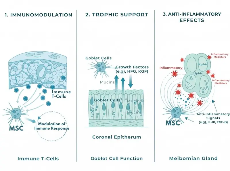
Mesenchymal stem cells (MSCs) — the type of cell used in our treatment program, found naturally in bone marrow, fat tissue, umbilical cord tissue, and several other locations in the body — have a well-documented ability to interact with and redirect immune cell behavior.
When introduced into a patient with active immune-driven lacrimal gland damage, MSCs release signaling proteins called cytokines (molecular messages between cells) that suppress the activity of the T-cells and B-cells responsible for infiltrating and destroying the gland tissue. At the same time, MSCs stimulate the growth of regulatory T-cells, often abbreviated as Tregs — a specialized immune cell population whose function is essentially to tell the rest of the immune system to stand down.
Think of it this way: in Sjögren’s syndrome-related dry eye syndrome, the immune system is treating the body’s own lacrimal gland as a foreign target. MSCs release signals that act like a ceasefire message — calming the attacking immune cells while amplifying the restraining ones. This does not cure the underlying autoimmune tendency. But by interrupting the active assault on gland tissue, it may allow what remains of the lacrimal gland’s secretory cell population to function more effectively, and in some patients, to partially recover function that had been suppressed rather than destroyed.
This mechanism is fundamentally different from what cyclosporine does at the ocular surface. Cyclosporine acts on surface cells. MSCs act on the immune cells generating the problem — closer to the source.
Supporting Ocular Surface Repair in Dry Eye Syndrome
The second mechanism addresses the damage that has already accumulated on the corneal and conjunctival surface.
MSCs secrete a broad range of growth factors — proteins that signal to neighboring cells to initiate repair processes. Among the most relevant for dry eye syndrome are growth factors that support the survival and function of corneal epithelial cells, promote the recovery of conjunctival goblet cells (the surface cells that produce the mucin layer which makes the tear film adhere to the eye), and help heal the fine disruption that chronic dry eye syndrome causes on the corneal surface over years of inflammation and exposure.
MSCs do not physically become corneal epithelial cells or goblet cells. What they do — through the growth factors and other signaling molecules they continuously release — is create a more favorable environment for the body’s own surface cells to survive, function, and slowly repair. In patients who respond, this translates to improved tear film stability and reduced staining on corneal surface assessment, which are the measures clinicians use to grade the severity of the surface damage in dry eye syndrome.
Protecting Meibomian Gland Function Against Dry Eye Syndrome Progression
The third mechanism relates specifically to the oil-producing Meibomian glands, whose dysfunction drives the evaporative form of dry eye syndrome.
Meibomian gland atrophy in MGD is driven in significant part by chronic periductal inflammation — inflammatory activity around the ducts and secretory cells of the gland. MSCs release anti-inflammatory signals that reduce this local inflammatory load. By calming periductal inflammation, stem cell therapy may slow the progressive atrophy of Meibomian gland tissue — protecting functioning gland capacity that still exists rather than regenerating capacity that has already been lost.
This is an important distinction. The mechanism here is protective, not regenerative in the classical sense. For patients with moderate MGD who still retain a meaningful proportion of functioning gland tissue, this slowing of progression is a clinically relevant benefit. For patients whose glands are almost entirely atrophied, there is less tissue for this protective mechanism to act on, and the anticipated benefit is smaller.
What Clinical Data Shows About Stem Cells and Dry Eye Syndrome
Outcome Data From Published Studies on Dry Eye Syndrome Stem Cell Treatment
The clinical evidence base for stem cell therapy specifically in dry eye syndrome is younger and less extensive than the evidence base for conditions such as osteoarthritis or ischemic heart failure, where randomized controlled trials have accumulated over fifteen or more years. This is a piece of honest context that is important to carry into any consideration of this treatment.
The most robust clinical data comes from trials examining MSC therapy in primary Sjögren’s syndrome, where dry eye is the most prevalent and often most disabling manifestation. A study published in Clinical Rheumatology examined the effects of allogeneic umbilical cord-derived MSC infusion in patients with primary Sjögren’s syndrome refractory to conventional treatment. At the 12-month assessment, participants showed statistically significant improvements in OSDI scores — the Ocular Surface Disease Index, a validated patient-reported outcome measure on a scale from 0 to 100, where scores above 33 indicate severe dry eye syndrome. A meaningful proportion of participants whose baseline OSDI scores were in the severe range showed reductions that moved them into the moderate range by 12 months. Schirmer test values — which measure aqueous tear production in millimeters per five minutes, with values below 5mm indicating severe deficiency — showed modest but statistically significant improvements compared to control groups.
A phase I/II trial examining subconjunctival injection of MSCs in patients with moderate-to-severe aqueous-deficient dry eye syndrome demonstrated a favorable safety profile across all participants and reported improvements in OSDI scores alongside measurable increases in goblet cell density on in-vivo confocal microscopy at six months. TBUT — tear break-up time, which measures how quickly the tear film becomes unstable after a blink (below 5 seconds is severe instability; above 10 seconds is normal) — showed improvements in a proportion of responding patients, suggesting some recovery of tear film stability at the surface level.
Studies using MSC-conditioned medium — a preparation containing the growth factors and signaling molecules secreted by MSCs, administered as topical drops — have demonstrated improvements in TBUT, reductions in corneal staining scores, and reduced surface inflammatory markers in controlled settings. These preparations represent a different delivery mechanism from systemic MSC infusion, but their results contribute to the evidence that MSC-derived signals have meaningful biological effects on the dry eye syndrome ocular surface.
What the Dry Eye Syndrome Clinical Data Does Not Yet Show
Precision about limitations is as important as precision about results.
There are no large-scale, multi-center Phase III randomized controlled trials comparing MSC therapy to best-available conventional treatment in a broad dry eye syndrome population with a pre-specified primary endpoint. The existing trials are generally small, single-center, and phase I/II in design — appropriate for establishing safety and early evidence of efficacy, but not yet sufficient to establish definitive clinical guidelines. This is an honest description of where the field stands for dry eye syndrome specifically, and it differs from conditions like osteoarthritis, where meta-analyses now synthesize data from dozens of randomized controlled trials.
The proportion of dry eye syndrome patients who achieve a clinically meaningful improvement in OSDI scores across published small trials is approximately 50 to 65 percent. A significant minority will see limited or no change, and we do not yet have reliable pre-treatment predictors for who will fall into which group.
Long-term durability beyond 12 to 18 months is not yet well characterized in the published dry eye syndrome literature. Whether the improvements observed at six to twelve months are sustained, whether repeat treatment is needed, and what the optimal dosing interval looks like over time are questions that active research is working to answer.
Autologous vs. Allogeneic: Comparing Stem Cell Sources for Dry Eye Syndrome
Why the Source of Stem Cells Matters in Dry Eye Syndrome Treatment
Not all stem cell therapies are equivalent, and the source of the cells has meaningful implications for what they can accomplish — particularly in a condition where the underlying driver is immune dysregulation.
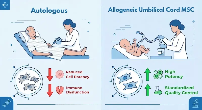
In Japan, the regulatory framework for regenerative medicine commonly supports autologous approaches: cells are taken from the patient’s own body, typically from bone marrow or fat tissue, processed in a laboratory, and reintroduced. The principal advantage is immunological — there is no rejection risk, because the cells are genetically the patient’s own.
The significant limitation for dry eye syndrome patients, particularly those with Sjögren’s syndrome, is that stem cell quality reflects the environment they came from. A patient in their late fifties with nine years of active autoimmune disease is asking cells that have lived inside that inflamed, immune-dysregulated body to perform immunomodulatory work. Published studies have documented that MSCs harvested from patients with systemic autoimmune conditions show reduced proliferative capacity, lower secretion of anti-inflammatory cytokines, and diminished Treg-stimulating activity compared to MSCs from healthy young donors. This is not a minor technical distinction. It has direct implications for how effectively those cells can modulate the immune attack on the lacrimal glands.
The Allogeneic Cord-Derived Approach Used in Malaysia for Dry Eye Syndrome
The approach we use in Malaysia relies on allogeneic MSCs derived from the Wharton’s jelly layer of donated umbilical cord tissue — from healthy newborn donors whose cells have not been exposed to decades of inflammation, drug treatment, or immune dysregulation.
Umbilical cord-derived MSCs show significantly higher proliferative activity, greater secretion of beneficial signaling molecules, and more potent immunomodulatory capacity than MSCs from adult donors or from patients with established inflammatory conditions. Because they express low levels of the surface markers that ordinarily trigger immune rejection, serious rejection reactions are uncommon — a finding consistently observed in large clinical trial populations across multiple MSC applications including rheumatoid arthritis, heart failure, and systemic autoimmune conditions.
The cells are prepared in standardized, quality-controlled batches. Cell viability and potency are verified before each treatment is administered. This consistency is not achievable with autologous preparations, where yield and functional quality vary with each individual patient and cannot be predicted in advance. For dry eye syndrome patients whose disease involves active immune-mediated gland destruction, the argument for using cells from an immunologically intact, biologically young donor rather than from the patient’s own compromised immune environment is directly supported by the mechanism through which the treatment is expected to work.
Who Is — and Is Not — a Good Candidate for Dry Eye Syndrome Stem Cell Therapy
Profiles That Respond Well to Dry Eye Syndrome Stem Cell Therapy
Stem cell therapy for dry eye syndrome is most likely to produce meaningful benefit in patients who meet a recognizable clinical profile.
The most appropriate candidates have moderate-to-severe dry eye syndrome confirmed by validated measures — OSDI scores in the moderate to severe range, Schirmer test values below 10mm/5min, and TBUT under 10 seconds — with an identifiable inflammatory or immune-mediated component. They have used preservative-free artificial tears consistently, trialed prescription anti-inflammatory drops for at least six months without achieving adequate relief, and have addressed Meibomian gland dysfunction through lid hygiene and, where appropriate, procedural treatment. Their disease has not yet progressed to complete end-stage atrophy of all secretory gland tissue — there is still functioning capacity remaining that could benefit from immune modulation and surface support.
Patients with primary Sjögren’s syndrome-related aqueous-deficient dry eye syndrome are the population most closely aligned with the biological mechanisms through which MSC therapy acts. If an inflammatory marker workup confirms active systemic autoimmune activity alongside lacrimal gland damage on imaging, the clinical rationale for MSC therapy is particularly coherent.
Patients with graft-versus-host disease affecting the eyes — a form of immune-mediated dry eye syndrome occurring after bone marrow transplantation — represent another profile in which the autoimmune mechanism is well-defined and the potential benefit of immune modulation is directly applicable.
When Dry Eye Syndrome Stem Cell Therapy Is Unlikely to Help
There are clinical situations where we advise against pursuing this treatment, either because benefit is unlikely or because a different approach is more appropriate.
Patients with complete end-stage Meibomian gland atrophy — where meibography or confocal microscopy shows that functional gland tissue has been almost entirely replaced by non-secreting material — have little remaining tissue for the protective mechanisms of MSC therapy to act on. In those cases, the more appropriate focus is on scleral lens fitting, moisture-chamber glasses, or other compensatory strategies that address near-total gland loss structurally rather than biologically.
Patients with dry eye syndrome caused primarily by eyelid malposition, incomplete eyelid closure (as in Bell’s palsy or thyroid eye disease), or neurotrophic corneal disease — where the surface lacks appropriate nerve signaling — have a structural or neurological cause that MSC therapy is not designed to address. These situations require targeted correction of the underlying anatomical or neurological problem.
Patients with mild dry eye syndrome that is adequately controlled on current treatment may not have enough to gain from this intervention to justify the time and cost. We will say this directly when it is our assessment.
Active ocular infection, active malignancy in the ocular region, uncontrolled systemic illness, or pregnancy are contraindications. Patients seeking rapid symptom relief — within days to weeks — should understand that the biological timeline of MSC therapy is incompatible with that need.
One Patient’s Experience With Dry Eye Syndrome and Stem Cell Treatment
After Years of Drops: Why She Pursued Dry Eye Syndrome Stem Cell Therapy
A woman in her mid-fifties had lived with primary Sjögren’s syndrome for eleven years. Her dry eye syndrome had been the most limiting aspect of the condition from the beginning, and over the years it had become the defining constraint of her working life. Her role required sustained computer use, and the progressive reduction in screen tolerance had begun to affect both the hours she could work and the quality of what she produced during them.
Over those eleven years she had used preservative-free artificial tears fifteen to twenty times daily, completed three separate courses of cyclosporine drops, trialed lifitegrast for eight months, had punctal plugs placed in all four puncta, and received four rounds of IPL treatment. Her OSDI score at the consultation before she came to us was 71. Her Schirmer test values — measured at her last ophthalmology appointment in Japan — were 2mm/5min in the right eye and 3mm/5min in the left. Her rheumatologist had noted a gradual upward trend in her inflammatory markers over the preceding eighteen months.
She was not coming to us with the expectation of a cure. What she asked directly was whether there was any biological basis for believing her lacrimal gland function could improve rather than continue to decline. After reviewing her imaging, inflammatory markers, and treatment history, we considered her an appropriate candidate for allogeneic MSC infusion on the basis of identified autoimmune activity, confirmed reduced but not absent residual tear production, and documented failure of all available conventional treatments. We were explicit before any treatment was arranged: we could not predict her individual response, the timeline for any change would be measured in months rather than weeks, and continuing all her existing dry eye management throughout and after the treatment was non-negotiable.
What Changed — and What Remained the Same — After Dry Eye Syndrome Treatment
The first six weeks produced no change she could detect — which she had been told to expect. Around week nine, she reported that her need for artificial tears had decreased from fifteen to twenty applications daily to nine or ten. The shift was gradual enough that she was not certain of it at first, then consistent enough that she was.
At the four-month assessment, she described being able to work at a computer for ninety-minute continuous intervals without drops — compared to twenty to thirty minutes before treatment. At the six-month follow-up, her OSDI score had reduced to 46, a 35 percent reduction from baseline, still in the clinically severe range but meaningfully different from where she had been. Her Schirmer test values had improved to 5mm/5min in the right eye and 6mm/5min in the left — modest by absolute measure, but a reversal of a trend that had been steadily declining for years. She remained on all her existing systemic Sjögren’s treatment and all her ocular management without modification.
Her description of the outcome: “I still have dry eye syndrome. I still use drops. But the ceiling on what I can do in a day has moved, and that matters more than I expected it to.”
Her improvement was real, partial, and took months to emerge. That is an accurate description of what stem cell therapy for dry eye syndrome can realistically provide — and it is the honest framing we offer every patient who considers this path.
※ This is an anonymized account based on actual clinical experience. Individual outcomes vary and cannot be guaranteed for any other patient.
What Recovery Looks Like After Dry Eye Syndrome Stem Cell Therapy
The physical recovery from MSC infusion is not demanding. There is no incision, no wound, and no significant restriction on daily activity beyond the first week. Most patients are comfortable travelling home within two to three days of treatment.
The most important aspect of the recovery period for dry eye syndrome specifically is patience with timeline. MSCs work through a biological process that unfolds over weeks to months. Patients who assess the treatment based on how they feel in the first two to four weeks are measuring before the relevant changes have had time to occur. The appropriate assessment window begins at six to eight weeks. The first meaningful signal that something is shifting is typically a gradual reduction in the daily frequency of artificial tear use — a change in management burden that often precedes any noticeable shift in symptom severity scores.
Honest Risks and Limitations of Dry Eye Syndrome Stem Cell Therapy
Side Effects and Safety Profile in Dry Eye Syndrome Stem Cell Treatment
The safety profile of allogeneic MSC therapy, across the published clinical literature in multiple inflammatory and autoimmune conditions, has been generally favorable. This is what the accumulated trial data shows, and it is important context for any individual patient’s decision.
The most commonly reported effects following infusion are mild and self-limiting. A low-grade fever in the 24 to 48 hours after infusion is reported by approximately 15 to 20 percent of patients — thought to reflect mild immune recognition of the donor cells rather than active infection — and resolves without specific treatment in the majority of cases. Transient fatigue and generalized muscle discomfort in the days after infusion are also commonly reported. These are not classified as serious adverse events in the published clinical literature.
Immune rejection of allogeneic MSCs has not emerged as a clinically significant problem in published trial data. MSCs express low levels of the surface proteins that typically trigger rejection responses and actively suppress the immune pathways that drive rejection. No cases of cell-related malignancy, tumor formation, or serious infection attributable to allogeneic MSC infusion from cord tissue have been reported in the peer-reviewed literature for this approach and cell type. This is a consistent finding across multiple conditions and multiple research groups.
For subconjunctival injection specifically, the risks include those associated with any ocular injection procedure: mild post-injection discomfort, temporary increased redness, and a low risk of infection managed through standard sterile technique. These are the same risk categories as any standard intraocular or periocular injection procedure.
What Stem Cells Cannot Do for Dry Eye Syndrome
Being direct about the limits of this treatment is as important as being direct about the evidence for its potential.
Stem cell therapy cannot regenerate Meibomian gland or lacrimal gland tissue that has undergone complete end-stage atrophy. When secretory cells have been entirely replaced by non-functioning fibrous tissue, the growth factors and immunomodulatory signals from MSCs do not have viable target tissue to work with. The treatment functions through existing residual tissue — it is a biological intervention, not a structural reconstruction.
It will not provide rapid relief. The biological timeline of MSC action is weeks to months, not days. Patients who require fast symptom control should pursue optimized conventional management rather than stem cell therapy as their primary response.
It will not eliminate the need for dry eye syndrome management. The realistic goal is a meaningful reduction in severity and in the daily burden of management — not independence from eye care. Patients will continue to need artificial tears, lid care, and regular ophthalmological review regardless of their response to this treatment.
Not every patient benefits. Based on published small trial data and our clinical experience, approximately 35 to 40 percent of dry eye syndrome patients treated with this approach may experience limited or no meaningful improvement. This number deserves to be stated clearly, and patients deserve to know it before making any decision.
Long-term outcome data beyond 18 months for dry eye syndrome specifically is limited. Whether improvements observed at six to twelve months are durable, whether repeat treatment is needed over time, and what optimal long-term dosing looks like are questions that ongoing research is working to address.
FAQ About Dry Eye Syndrome Stem Cell Treatment
Cyclosporine and lifitegrast work on the surface of the eye, reducing T-cell activity on the corneal and conjunctival surface. They do not reach the lacrimal glands and cannot interrupt the immune infiltration destroying those glands from the inside. MSC therapy works at the level of the immune cells themselves — the T-cells and B-cells driving gland damage — rather than at the ocular surface where the effects of that damage are visible. The two approaches are not mutually exclusive; most patients continue existing eye drops alongside and after MSC therapy.
In most cases, yes. The aim of this treatment is a meaningful reduction in symptom severity and in the frequency of management required — not the elimination of dry eye syndrome management altogether. Patients who respond typically describe needing drops less frequently and tolerating visual tasks better, but ongoing management remains part of the picture.
The first measurable changes typically emerge between six and ten weeks after treatment. Objective changes in Schirmer test values or TBUT, where they occur, are generally visible at the three to six month assessment. The first subjective signal most patients notice is a gradual reduction in how often they need to use drops — a change in management burden that often precedes any clear shift in severity scores. Assessing the treatment based on the first month produces an inaccurate picture.
Sjögren’s syndrome-related dry eye is one of the presentations for which the biological rationale for MSC therapy is most directly coherent, and it is the patient population in which the most relevant published clinical trial data has been generated. The immune-mediated destruction of lacrimal gland tissue in Sjögren’s syndrome is precisely what MSC immunomodulation is designed to interrupt. Whether a given patient with Sjögren’s syndrome is an appropriate candidate depends on the degree of residual gland function, current disease activity, and other factors assessed in the pre-treatment review.
This is a self-funded private medical procedure and is not covered by health insurance in Japan, Malaysia, or most other countries. We recommend verifying the specifics of your own policy, but coverage for this type of treatment is not currently available in our experience.
If You Have Reached the End of the Conventional Road With Dry Eye Syndrome
Living with dry eye syndrome that has outgrown conventional treatment is a particular kind of exhaustion — the quiet daily negotiation of managing a condition that progressively limits more. If you have reached the point where your current treatment keeps you stable but does not move you forward, and you are asking whether there is a biological option that could address what is happening at the level of the glands, we understand why that question is worth pursuing honestly.
We offer free online consultations for exactly this kind of conversation. Bring your most recent assessments — OSDI scores, Schirmer test values, a history of what you have tried — and we will review your case and tell you straightforwardly whether your profile falls within the range where this treatment has shown evidence of benefit. If it does not, we will tell you that too. You are not committing to anything other than the conversation itself.
References
- Craig JP, et al. TFOS DEWS II Report Executive Summary. Ocular Surface. 2017;15(4):802–812. https://doi.org/10.1016/j.jtos.2017.08.003
- Bron AJ, et al. TFOS DEWS II pathophysiology report. Ocular Surface. 2017;15(3):438–510. https://doi.org/10.1016/j.jtos.2017.05.011
- Jones L, et al. TFOS DEWS II Management and Therapy Report. Ocular Surface. 2017;15(3):575–628. https://doi.org/10.1016/j.jtos.2017.05.006
- Liang J, et al. Allogeneic mesenchymal stem cells transplantation in patients with refractory systemic lupus erythematosus: a pilot clinical study. Ann Rheum Dis. 2010;69(8):1423–1429. https://doi.org/10.1136/ard.2009.123463
- Wang D, et al. Safety and preliminary efficacy of umbilical cord-derived mesenchymal stem cells in primary Sjögren’s syndrome. Stem Cell Translational Medicine. 2016;5(12):1745–1754. https://doi.org/10.5966/sctm.2015-0378
- Xu J, et al. Allogeneic mesenchymal stem cell treatment alleviates experimental and clinical Sjögren syndrome. Blood. 2012;120(15):3142–3151. https://doi.org/10.1182/blood-2011-11-391144
- Weng J, et al. Mesenchymal stem cell-conditioned medium promotes corneal epithelial wound healing. Stem Cells International. 2017;2017:1–10. https://doi.org/10.1155/2017/3850264
- Pflugfelder SC, de Paiva CS. The Pathophysiology of Dry Eye Disease: What We Know and Future Directions for Research. Ophthalmology. 2017;124(11S):S4–S13. https://doi.org/10.1016/j.ophtha.2017.07.010
- Mead B, Tomarev S. Bone marrow-derived MSC-secreted exosomes promote survival of retinal ganglion cells through miRNA-dependent mechanisms. Stem Cells Translational Medicine. 2017;6(4):1273–1285. https://doi.org/10.1002/sctm.16-0428
- Le Blanc K, Ringdén O. Immunomodulation by mesenchymal stem cells and clinical experience. J Intern Med. 2007;262(5):509–525. https://doi.org/10.1111/j.1365-2796.2007.01844.x