
Post-Stroke Sequelae Stem Cell Therapy | Can Recovery Continue After Rehab Plateaus?
The coffee cup in your left hand trembles in a way it never used to. Your spouse has learned to walk a half-step slower when the two of you go to the corner store together. The joke you wanted to tell last night kept slipping out of reach, the words refusing to arrive in the order they used to.
If you are living with the aftermath of a stroke — or accompanying someone who is — you are part of a population larger than most people realize. According to the Global Burden of Disease Study, more than 101 million people worldwide are living with the residual effects of stroke. Roughly half of all stroke survivors carry permanent neurological deficits of some kind: weakness or paralysis on one side of the body, difficulty with speech or comprehension, problems with balance, fatigue, or cognitive changes that the people closest to them notice before the medical team does.
For most survivors, the months following the stroke follow a recognizable arc. Intensive inpatient rehabilitation gives way to outpatient therapy. Outpatient therapy gives way to home exercise. Improvement comes in measurable steps for a while, and the milestones — first standing, first sentence, first day without the cane — accumulate. Then, somewhere between six months and a year after the stroke, the improvements slow. Therapists call it the plateau. It is the point at which standard rehabilitation can no longer reliably produce further functional gains. The deficits that remain at the plateau are, in most cases, the deficits the patient has been told — gently or directly — to learn to live with.
This article is written for people who have reached that plateau, or who are watching a parent, spouse, or sibling reach it, and who are asking whether anything more can be done at the biological level — beyond more therapy, beyond more medication.
We will be direct about what stem cell therapy for post-stroke sequelae can and cannot do. The clinical evidence is meaningful but incomplete. Some patients show measurable functional improvement; others do not. Our clinic in Malaysia has worked in regenerative medicine for over seven years, with more than 200 patients treated across multiple conditions. The aim here is to give you what we believe you need to make an informed decision — the numbers from the trials, the patient profiles that tend to respond, and the situations in which we would honestly advise against this treatment.
Table of Contents
- Why Post-Stroke Sequelae Don't Resolve on Their Own
- Why Conventional Post-Stroke Treatment Reaches a Ceiling
- How Stem Cell Therapy Targets Post-Stroke Sequelae
- What Clinical Trials Show for Post-Stroke Sequelae Stem Cell Therapy
- Who Is — and Is Not — a Good Candidate for Post-Stroke Sequelae Stem Cell Therapy
- A Patient's Experience with Post-Stroke Sequelae Stem Cell Therapy
- Honest Risks and Limitations of Post-Stroke Sequelae Stem Cell Therapy
- FAQ About Post-Stroke Sequelae Stem Cell Therapy
- Considering Your Options for Post-Stroke Sequelae
- References
Why Post-Stroke Sequelae Don’t Resolve on Their Own
The Brain’s Limited Repair Mechanisms After Stroke
A stroke is, at its core, a sudden interruption of blood supply to part of the brain. In the most common form — ischemic stroke, accounting for roughly 85 percent of cases worldwide — a blood vessel becomes blocked, and the brain tissue downstream of the blockage is deprived of oxygen and glucose. Within minutes, neurons in the core of the affected area begin to die. Around this core sits a surrounding zone of tissue, called the penumbra, that is critically under-supplied but not yet dead. Whether the penumbra survives or joins the core in death determines much of what the long-term deficits will look like.
The brain has historically been described as one of the least regenerative tissues in the body. That picture has become slightly more nuanced over the past two decades — adult neurogenesis (the formation of new neurons) does occur in certain limited regions, and the brain demonstrates significant plasticity in how surviving regions can take over functions from damaged ones. But the fundamental fact remains: once neurons in the stroke core die, they do not come back. The lost tissue is replaced by a glial scar — a dense fibrotic structure formed by support cells called astrocytes — and that scar does not transmit signals or perform any of the functions of working neural tissue.
What recovery actually consists of, in the standard rehabilitation model, is reorganization. Surviving regions of the brain, often in the opposite hemisphere or in nearby intact areas, gradually take on some of the work the damaged region used to do. New connections form between neurons that had previously been less involved in that function. Repeated, structured practice — the work of rehabilitation — drives this reorganization. It is a real and remarkable process, but it has limits.
Why Recovery Plateaus After 6 to 12 Months
The recovery curve after stroke is steepest in the first three months, continues at a slower rate through six months, and tends to flatten substantially between six and twelve months. Multiple longitudinal studies have documented this pattern. By the one-year mark, the deficits that remain are largely the deficits the patient will live with going forward — unless something changes the underlying biology.
Several things drive the plateau. The brain’s plasticity is greatest in the early period after injury, when growth factors and other repair signals are most actively produced. Over time, those signals diminish. The peri-infarct tissue — the region surrounding the dead core — settles into a chronic state of low-grade inflammation that, paradoxically, both reflects ongoing injury and inhibits further repair. The glial scar that walls off the damaged tissue, while protective in the acute phase, also acts as a physical and biochemical barrier to new connections forming across it.
This is the biological situation that any treatment claiming to produce further recovery after the plateau must address. Rehabilitation, which works by driving plasticity in surviving tissue, can continue to help — but it works against a slowly closing biological window. Stem cell therapy attempts to reopen that window, at least partially, by changing the chemical environment in which the surviving tissue operates.
Why Conventional Post-Stroke Treatment Reaches a Ceiling
What Rehabilitation Achieves — and Where It Ends
Rehabilitation is not a placeholder treatment, and we want to be clear about that before describing its limits. Structured physical therapy, occupational therapy, and speech and language therapy after stroke have been shown in multiple controlled trials to produce meaningful improvements in function, particularly in the first three to six months. For many patients, the difference between aggressive early rehabilitation and minimal early rehabilitation is the difference between independence and dependence in daily life a year later.
The challenge is what happens after the plateau. Trial after trial of various intensified rehabilitation protocols — extended hours, robotic-assisted training, constraint-induced movement therapy, repetitive transcranial magnetic stimulation paired with practice — have shown that additional gains are possible in chronic stroke, but they are typically modest. The plateau is not an absolute wall, but the slope of further improvement under standard approaches is shallow.
For a patient who is twelve or eighteen months out from their stroke and still cannot reliably grip a coffee cup with their affected hand, or whose speech is still effortful, or who still loses balance walking on uneven ground, the conversation with the rehabilitation team tends to shift. The focus moves from restoring function to compensating for what is missing — different techniques for getting dressed, different strategies for communication, modifications to the home environment. These compensations are important and valuable. They are not, however, the recovery the patient was hoping for.
The Limits of Medications and Devices for Chronic Stroke Deficits
The medications used after stroke are primarily directed at preventing a second stroke, not at improving recovery from the first. Antiplatelet agents, anticoagulants in certain types of stroke, blood pressure medications, statins (cholesterol-lowering drugs) — these are essential for secondary prevention, and most stroke patients will be on some combination of them indefinitely. None of them, however, directly drives neurological recovery.
A handful of medications have been studied for their potential to enhance post-stroke recovery — selective serotonin reuptake inhibitors (a class of antidepressant), particularly fluoxetine, have shown some signal in earlier trials but more recent and larger trials have produced mixed results. Modafinil has been studied for post-stroke fatigue with limited evidence. None of these agents has emerged as a reliable tool for producing further functional improvement after the plateau.
Devices represent a more recent frontier. Functional electrical stimulation, exoskeletons, brain-computer interfaces, and various forms of non-invasive brain stimulation have been studied with results that are often promising in research settings but slower to translate into widespread clinical benefit. For most patients past the rehabilitation plateau, what is realistically available outside of clinical trials remains rehabilitation, secondary prevention medications, and adaptive equipment.
It is this gap — between what the patient has already received and what they are still looking for — that drives the question of whether stem cell therapy might offer something different.
How Stem Cell Therapy Targets Post-Stroke Sequelae
Three Mechanisms in the Post-Stroke Brain
Mesenchymal stem cells, or MSCs, do not work the way many people initially imagine. Early in the history of stem cell research, there was significant hope that infused cells would migrate to damaged tissue, transform into the missing cell type — in this case, new neurons — and physically rebuild what had been lost. That expectation has not held up under closer scientific scrutiny. MSCs introduced into the body after stroke do not reliably differentiate into functioning neurons that integrate into existing neural circuits.
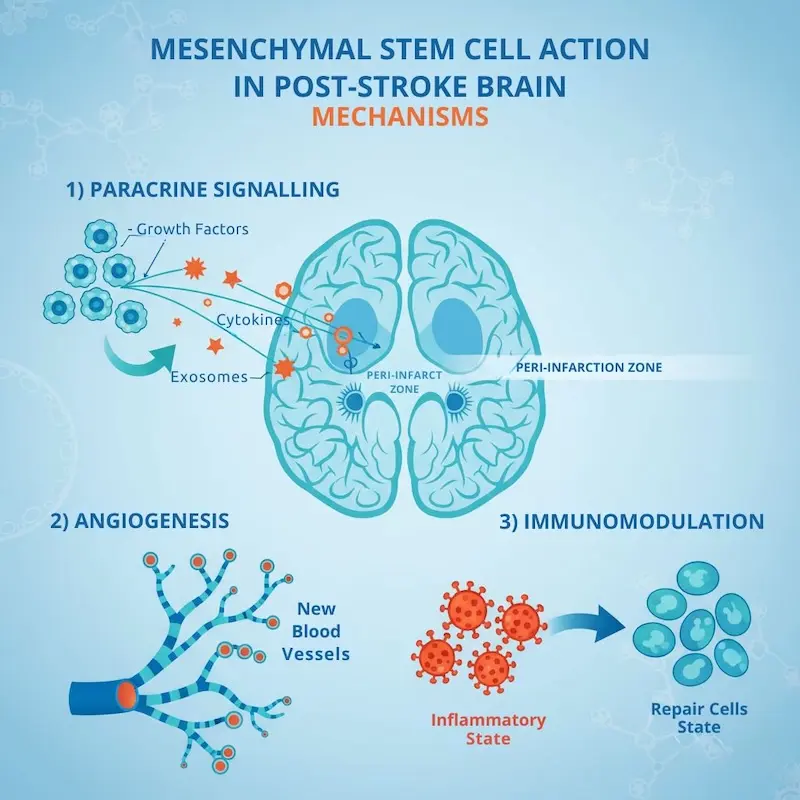
What the accumulated evidence suggests instead is that MSCs work indirectly, through three primary mechanisms that together change the environment surrounding the injured tissue.
The first is paracrine signaling. MSCs secrete a wide range of molecules — growth factors, cytokines (chemical messengers between cells), and extracellular vesicles called exosomes — that communicate with neighboring cells. In the post-stroke brain, these signals appear to support the survival of neurons in the peri-infarct zone, encourage the sprouting of new connections from healthy neurons into damaged regions, and prolong the brain’s own resident repair processes that would otherwise have faded with time.
The second is angiogenesis — the formation of new small blood vessels. Regions of the brain that have been chronically under-perfused since the stroke can remain alive but severely under-functioning, similar to the way under-supplied heart muscle behaves in chronic heart failure, where the same biological process is studied as a route to recovering function in tissue that has been struggling for years.
The third is immunomodulation. The post-stroke brain remains in a state of low-grade chronic inflammation for months and sometimes years after the acute event. This inflammation is not benign — it actively interferes with the brain’s repair processes and contributes to the secondary loss of neurons over time. MSCs have a well-documented ability to shift immune cells from a destructive, inflammatory state to a reparative state, both within the brain itself and at the level of the systemic immune system that influences the brain through the bloodstream. The same immunomodulatory capacity is the basis for using MSCs in autoimmune conditions such as rheumatoid arthritis, where the inflammatory pathways involved overlap meaningfully with those active in chronic post-stroke brain tissue.
The combined effect, when these mechanisms work as intended, is an environment in which surviving neural tissue can perform better, in which the brain’s own plasticity processes have more biological support, and in which the chronic inflammatory signaling that has been suppressing recovery is reduced.
Allogeneic Umbilical Cord MSCs vs. Autologous Cells for Stroke
Stem cell therapy comes in two basic structures: autologous, using cells from the patient’s own body, and allogeneic, using cells from a healthy donor.
In Japan, the current regulatory framework supports autologous treatment in most contexts. Cells are typically harvested from the patient’s own bone marrow or adipose (fat) tissue, processed in a laboratory, and returned. The advantage is that there is no immune compatibility concern, since the cells originated in the patient. The disadvantage, particularly for stroke patients, is that the cells reflect the patient’s age and health status. A 70-year-old patient who has had a stroke, who likely has some combination of hypertension, diabetes, vascular disease, and the systemic effects of years of these conditions, is harvesting cells from an environment that has shaped how those cells function. Multiple studies have suggested that MSCs from older donors and from donors with cardiovascular disease show measurably reduced proliferative capacity and reduced secretion of beneficial signaling molecules.
The treatment we offer in Malaysia uses allogeneic umbilical cord-derived MSCs. These cells are sourced from ethically donated umbilical cord tissue (specifically the Wharton’s jelly layer) collected at the time of healthy newborn deliveries with full donor consent. They are biologically young, highly active in their signaling output, and produced in standardized batches with consistent quality testing. Umbilical cord MSCs express low levels of the surface proteins that ordinarily trigger immune rejection, which is why allogeneic infusions of these cells have shown consistently low rates of rejection-related complications in published trials.
The choice of cell source is not merely technical. For a stroke patient in their sixties or seventies, the difference between cells from a healthy newborn donor and cells from their own aged, vascular-disease-affected body has direct consequences for how much signaling activity reaches the brain.
What Clinical Trials Show for Post-Stroke Sequelae Stem Cell Therapy
Major Stroke Stem Cell Trials and Their Numbers
The clinical trial landscape for stroke stem cell therapy has grown substantially over the past fifteen years. We will summarize what the more methodologically sound trials have shown, with specific numbers, because vague summaries do not help patients make informed decisions.
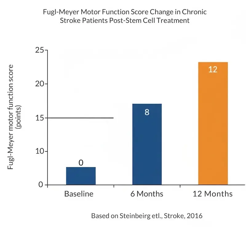
The Stanford SB623 study, published by Steinberg and colleagues in the journal Stroke in 2016, was an early and influential phase 1/2a trial. It enrolled 18 patients with chronic stroke — between six months and three years post-stroke — who had stable motor deficits. The cells used were modified allogeneic bone marrow-derived MSCs, delivered through a single stereotactic neurosurgical injection into the brain region adjacent to the stroke. At twelve months, patients showed statistically significant improvements on multiple standardized outcome scales: the European Stroke Scale, the National Institutes of Health Stroke Scale, and most notably the Fugl-Meyer motor function scale (a standardized measure of motor recovery), where the mean improvement was 11.4 points. To place that in context, a change of 4 to 6 points on the Fugl-Meyer is generally considered clinically meaningful. Treatment-related adverse events were primarily linked to the surgical delivery procedure rather than to the cells.
The PISCES program — Pilot Investigation of Stem Cells in Stroke — used a different cell type, a conditionally immortalized human neural stem cell line called CTX0E03, also delivered surgically. The PISCES-II trial, published by Muir and colleagues in 2020, enrolled 23 patients with chronic upper limb hemiparesis (weakness of the arm and hand on one side) between six months and five years post-stroke. The primary endpoint was an improvement of two or more points on the Action Research Arm Test at three months — a meaningful change in functional use of the affected arm. Of the 23 patients treated, 12 met this threshold, representing 52 percent of the trial population.
A 2019 trial published in Stroke by Levy and colleagues took a different approach: intravenous infusion of allogeneic bone marrow-derived MSCs in chronic stroke patients, rather than surgical implantation into the brain. This trial enrolled 36 patients and assessed safety and preliminary efficacy. The infusion was well tolerated, with no serious treatment-related adverse events reported. Functional improvements on the Barthel Index (a measure of independence in daily activities) and modified Rankin Scale (a measure of global disability) were observed in the treated group.
The MASTERS trial, published by Hess and colleagues in The Lancet Neurology in 2017, took yet another approach: intravenous infusion of MultiStem (a related allogeneic cell product) in patients within 24 to 48 hours of acute ischemic stroke, with a focus on early intervention rather than chronic deficits. The primary efficacy endpoint was not met in the overall trial population, though pre-specified subgroup analyses suggested benefit in patients treated earliest. The follow-up program has continued to study this question.
What Realistic Improvement Looks Like After Treatment
We want to translate these trial numbers into what realistic improvement actually looks like for a patient, because trial endpoints often obscure the human meaning of the data.
A clinically meaningful Fugl-Meyer improvement might mean the difference between a hand that cannot grip anything and a hand that can hold a phone or a coffee cup, even if the fine motor control of individual fingers remains limited. A two-point improvement on the Action Research Arm Test might mean being able to reach a shelf, or pick up a pen, that the patient previously could not. A change of one point on the modified Rankin Scale might mean walking with a cane rather than a walker, or walking unassisted rather than with a cane.
These changes are real and they matter — but they are not, in most cases, full restoration of pre-stroke function. The published data does not support the idea that stem cell therapy can return a chronic stroke patient to their pre-stroke baseline. What it suggests, in the patients who respond, is meaningful incremental improvement layered onto whatever recovery has already occurred.
It is also important to note that not every patient responds. Across the published trials, response rates have varied widely depending on patient selection, cell source, delivery method, and the specific endpoint being measured. In the PISCES-II trial, 52 percent of patients met the primary functional improvement threshold — which is encouraging but also means that close to half did not. We do not currently have a reliable way to predict in advance which individual patients will fall into which group, though several factors discussed below appear to influence the odds.
Who Is — and Is Not — a Good Candidate for Post-Stroke Sequelae Stem Cell Therapy
Profiles That Tend to Respond to Stroke Stem Cell Therapy
Based on the available trial data and the patient selection criteria those trials used, several characteristics appear to predict a higher likelihood of meaningful response.
Patients who are six months to roughly three years post-stroke, who have entered the rehabilitation plateau but are not many years out from the event, tend to fit the profile of trial responders. The biology of the post-stroke brain remains more responsive to repair signals in this window than years after the event, although meaningful improvements have been reported in some patients even five years post-stroke.
Ischemic stroke patients with deficits primarily related to motor function — weakness or paralysis on one side — have been the most studied population. Other deficit types such as aphasia, cognitive impairment, and sensory deficits have less specific trial data, though many trials have reported improvements across multiple deficit categories alongside motor improvements.
Patients whose stroke was a single, identifiable event with a defined area of damage tend to be better candidates than those with multiple infarcts (small strokes) scattered across the brain. The presence of viable peri-infarct tissue, identifiable on MRI, is a positive prognostic factor. This is the tissue that the mechanisms of MSC therapy are most likely to support.
Stable medical condition at the time of treatment is essential. The patient should be on optimized secondary prevention medications, hemodynamically stable, without active infection, and not in a phase of acute medical decompensation. The cardiovascular risk factors that contributed to the original stroke overlap significantly with the conditions managed in chronic heart failure care, and controlling them remains essential whether or not stem cell therapy is pursued.
Finally — and this matters — patients who continue active rehabilitation alongside or after stem cell treatment appear to do better than those who treat stem cell therapy as a substitute for ongoing therapy. The biological signals from MSCs and the targeted neural retraining from rehabilitation work on overlapping mechanisms; the combination tends to produce more than either alone.
When Stem Cell Therapy Is Unlikely to Help Post-Stroke Sequelae
There are situations where we will not accept a patient for this treatment, or where we will tell them honestly that the likelihood of benefit is low.
End-stage deficits with no remaining viable tissue in the affected region are unlikely to respond to MSC therapy. If imaging shows the entire region responsible for the affected function has been replaced by glial scar and there is no preserved peri-infarct tissue, the biological substrate for the treatment’s mechanisms is absent.
Hemorrhagic stroke is biologically different from ischemic stroke, and the clinical trial evidence is substantially more limited for stem cell therapy in this setting. We assess hemorrhagic stroke cases individually but with greater caution about likely benefit.
Patients with active malignancy should not undergo MSC therapy, due to the theoretical concern that the growth-supportive signaling profile of MSCs could affect tumor behavior. Active systemic infection is also a contraindication.
Patients who are very recently post-stroke — within the first three months — are typically still in the most productive phase of standard rehabilitation, and the conversation about stem cell therapy is usually premature. The question becomes more relevant when conventional approaches have begun to plateau.
Patients seeking full restoration of pre-stroke function should understand clearly that this is not what the current evidence supports. We discuss expectations directly in initial consultations and decline to proceed when we believe the patient is operating with expectations the treatment cannot meet.
Recovery Timeline After Post-Stroke Sequelae Stem Cell Therapy
The recovery timeline after stem cell therapy is fundamentally different from what most patients are accustomed to from acute medical interventions. There is no rapid effect to expect in the first days or weeks. The biological processes that the cells initiate — angiogenesis, paracrine signaling, immunomodulation — operate on a timescale of weeks to months rather than hours to days.
Most patients who respond begin to notice changes between weeks 8 and 16 post-treatment. The earliest changes patients commonly report are not in the most affected limb but in general energy levels, fatigue, sleep quality, and cognitive clarity. Motor function changes, when they occur, tend to emerge slightly later — typically between months 3 and 6, with continuing gradual evolution through month 12.
We strongly encourage patients to continue or increase their rehabilitation activities during this period. The window of enhanced biological responsiveness is also a window of opportunity for the kind of targeted retraining that physiotherapy, occupational therapy, and speech and language therapy provide. Patients who pause their rehabilitation after stem cell treatment, hoping the cells will do the work for them, generally see less benefit than those who use the treatment as a foundation for renewed therapeutic effort.
A Patient’s Experience with Post-Stroke Sequelae Stem Cell Therapy
The Decision Point: Eighteen Months After a Middle Cerebral Artery Stroke
A man in his early sixties had a right middle cerebral artery ischemic stroke. He survived the acute event, received timely thrombolytic treatment, and entered an intensive rehabilitation program within days. Over the following twelve months he made significant progress — from initially unable to move his left side, to walking with a cane, to managing most activities of daily living with adaptive techniques. At fifteen months post-stroke his rehabilitation team described his progress as having plateaued. His left hand remained weak and clumsy, capable of stabilizing objects but not of fine manipulation. His gait remained asymmetric. He had returned to work in a reduced capacity but described the cognitive load of compensating as exhausting.
After researching the published trials and consulting with our team, he elected to proceed with allogeneic umbilical cord MSC therapy in Malaysia in early 2025, at eighteen months post-stroke. We were direct with him in the pre-treatment consultation: we could not predict his individual response, the treatment was unlikely to restore pre-stroke function, and any improvements he experienced would likely emerge gradually over months rather than weeks.
How Recovery Unfolded Over Twelve Months
In the first six weeks following treatment he noticed nothing he could clearly attribute to the therapy. He described this period later as the part that tested his patience most. By the third month he reported that the fatigue he had carried since the stroke was noticeably reduced — not eliminated, but enough to change how he ended his work days. He was sleeping better.
By the sixth month his physiotherapist documented measurable improvements on the Fugl-Meyer scale that had not been seen in the previous twelve months of consistent therapy. He could button a shirt — slowly — without help. His gait analysis showed improvements in stride symmetry. At twelve months post-treatment his improvements had stabilized but not reversed, and he had reduced his use of the cane to long walks and uneven terrain.
He continues active physiotherapy and remains on all of his secondary prevention medications. He describes the change as meaningful in daily life rather than dramatic — not a return to who he was before the stroke, but a measurable widening of what he can do.
※ This is an anonymized account shared with the patient’s consent. Individual outcomes vary. This experience does not represent what all patients will experience.
Honest Risks and Limitations of Post-Stroke Sequelae Stem Cell Therapy
Known Side Effects and Safety Data
The safety profile of intravenous allogeneic MSC therapy across published clinical trials has generally been favorable. The most common reported effects are mild and self-limiting: a low-grade fever in the 24 to 48 hours after infusion, transient fatigue, occasional headache, and mild flu-like symptoms. These typically resolve without intervention and are thought to reflect the immune system’s recognition of the donor cells rather than any active infection.
Serious adverse events directly attributable to the cells themselves have been uncommon across published stroke stem cell trials. The MASTERS trial, which enrolled patients shortly after acute stroke, found that intravenous MSC infusion did not increase the rate of adverse events compared to placebo. The Steinberg 2016 SB623 trial reported some adverse events related to the neurosurgical delivery method — headaches, transient neurological symptoms — rather than to the cells themselves; intravenous delivery, as used in our protocol, avoids these surgical risks.
Long-term safety data beyond two to three years remains more limited, reflecting the relative youth of the field at clinical scale. We disclose this to every patient before treatment, and we participate in long-term follow-up of our own patients to contribute to the broader understanding of post-treatment outcomes.
What Stem Cell Therapy Cannot Do for Post-Stroke Sequelae
This section matters as much as any other.
Stem cell therapy cannot rebuild brain tissue that has been destroyed by stroke. The neurons in the stroke core do not come back. What MSCs may support is the function of surviving tissue in and around the damaged region — they do not regenerate lost neural circuitry.
It cannot restore pre-stroke function in patients with substantial chronic deficits. The most realistic framing is that of meaningful incremental improvement on top of existing recovery, not a return to baseline.
It cannot replace ongoing rehabilitation. Patients who continue active physical, occupational, and speech therapy after treatment do better than those who do not. This pattern is consistent across the trial data and our clinical experience.
It cannot guarantee improvement. Based on the published trial data and our experience over seven years, a meaningful proportion of patients — somewhere between 40 and 60 percent depending on patient profile — do not experience a clinically significant change following treatment. We cannot predict with certainty which patients will respond. Patients deserve to know this number before making any decision.
It cannot replace ongoing secondary prevention. Stroke patients remain at elevated risk of a second stroke, and the medications, lifestyle interventions, and cardiovascular monitoring that reduce this risk remain essential whether or not stem cell therapy is pursued. The cardiovascular risk factors that contributed to the first stroke — hypertension, diabetes, atrial fibrillation, atherosclerosis — must continue to be managed actively, alongside any regenerative treatment.
FAQ About Post-Stroke Sequelae Stem Cell Therapy
The published clinical trials have primarily enrolled patients between six months and three years post-stroke, with some trials including patients up to five years out. The biological window for response is widest in the earlier part of this range — between six months and roughly two years — because the surrounding tissue still retains greater responsiveness to the repair signals that mesenchymal stem cells provide. That said, meaningful improvements have been documented in some patients further out from their stroke, and we assess each case individually rather than applying a strict time cutoff. Patients within the first three months of their stroke are typically still in the most productive phase of standard rehabilitation, and we usually recommend completing that phase before considering stem cell therapy. The shorter answer is: the window is wider than many patients assume, but it does narrow with time, and earlier within the chronic phase is generally more favorable than later.
It is important to be honest about this. Stem cell therapy does not rebuild brain tissue that has been destroyed, and it cannot restore neurons that have died in the stroke core. What the clinical trial data suggests it may do — in patients who respond — is improve the function of surviving tissue around the damaged region, support the brain’s existing plasticity processes, and produce measurable functional gains that further rehabilitation alone has not been able to achieve. For a patient with complete paralysis from a large stroke that destroyed the entire motor region, the realistic likelihood of regaining purposeful movement is low. For a patient with partial weakness who has plateaued, the published trials suggest that meaningful incremental improvement — better grip, easier gait, more reliable hand function — is possible in a significant proportion of cases. The accurate framing is improvement rather than reversal.
Rehabilitation and stem cell therapy work through different but complementary mechanisms. Rehabilitation drives recovery through repeated, structured practice that triggers reorganization in surviving brain regions — it works directly on the wiring level. Stem cell therapy works on the biological environment in which that wiring sits: reducing the chronic low-grade inflammation that has settled into the post-stroke brain, supporting the survival of struggling neurons in the peri-infarct region, and promoting the formation of new small blood vessels in under-perfused tissue. Neither replaces the other. The pattern we consistently see, both in published trials and in our own follow-up, is that patients who continue active rehabilitation alongside or after stem cell treatment experience more functional gain than those who stop rehabilitation in the expectation that the cells will do the work for them. The treatment is most accurately understood as creating better biological conditions for rehabilitation to continue working, rather than as a substitute for it.
The published trials have follow-up periods of generally one to three years, and within that window, improvements observed at the six-to-twelve-month mark have tended to be maintained rather than regressing. The Steinberg SB623 trial documented sustained motor function improvements at twelve months, and follow-up reports from several of the surgical and intravenous trials have continued to show stable gains in responding patients over multiple years. Longer-term data beyond three years remains limited, which is an honest gap in the current evidence. What we can say is that the gains, when they occur, do not appear to be transient effects that fade after weeks or months — they reflect actual changes in how the brain’s surviving tissue is functioning. Whether some patients might benefit from a second treatment course years later is a question the field has not yet answered with controlled data; we discuss this with patients individually rather than recommending a fixed schedule.
Yes, and we strongly advise that patients continue all of their existing secondary prevention medications without interruption. The risk of a second stroke remains real, and the medications that reduce that risk — antiplatelet agents, anticoagulants, blood pressure medications, statins, and others depending on the underlying cause of the original stroke — are essential whether or not stem cell therapy is pursued. We do not ask any patient to stop or alter their cardiovascular medications before, during, or after MSC treatment. Any decisions about medication adjustment should be made by the patient’s neurologist or primary care physician based on overall clinical status, not in connection with stem cell therapy. The treatment is designed to work alongside established stroke care, not as a replacement for it.
Considering Your Options for Post-Stroke Sequelae
If you have read this far, you are most likely either someone navigating the post-stroke plateau yourself or accompanying someone you love through it. We understand that this is a position in which decisions can feel heavy, and information from different sources rarely lines up cleanly.
We offer free online consultations specifically for cases like this. Bring the imaging, the rehabilitation history, and the questions. We will tell you honestly whether your situation fits the profile of patients the available evidence supports — and if it does not, we will tell you that as well. There is no obligation to proceed, and no decision needs to be made on the first call.
References
- GBD 2019 Stroke Collaborators. “Global, regional, and national burden of stroke and its risk factors, 1990–2019: a systematic analysis for the Global Burden of Disease Study 2019.” The Lancet Neurology. 2021;20(10):795–820. https://doi.org/10.1016/S1474-4422(21)00252-0
- Steinberg GK, Kondziolka D, Wechsler LR, et al. “Clinical Outcomes of Transplanted Modified Bone Marrow–Derived Mesenchymal Stem Cells in Stroke: A Phase 1/2a Study.” Stroke. 2016;47(7):1817–1824. https://doi.org/10.1161/STROKEAHA.116.012995
- Kalladka D, Sinden J, Pollock K, et al. “Human neural stem cells in patients with chronic ischaemic stroke (PISCES): a phase 1, first-in-man study.” The Lancet. 2016;388(10046):787–796. https://doi.org/10.1016/S0140-6736(16)30513-X
- Muir KW, Bulters D, Willmot M, et al. “Intracerebral implantation of human neural stem cells and motor recovery after stroke: multicentre prospective single-arm study (PISCES-2).” Journal of Neurology, Neurosurgery and Psychiatry. 2020;91(4):396–401. https://doi.org/10.1136/jnnp-2019-322515
- Hess DC, Wechsler LR, Clark WM, et al. “Safety and efficacy of multipotent adult progenitor cells in acute ischaemic stroke (MASTERS): a randomised, double-blind, placebo-controlled, phase 2 trial.” The Lancet Neurology. 2017;16(5):360–368. https://doi.org/10.1016/S1474-4422(17)30046-7
- Levy ML, Crawford JR, Dib N, Verkh L, Tankovich N, Cramer SC. “Phase I/II Study of Safety and Preliminary Efficacy of Intravenous Allogeneic Mesenchymal Stem Cells in Chronic Stroke.” Stroke. 2019;50(10):2835–2841. https://doi.org/10.1161/STROKEAHA.119.026318
- Savitz SI, Cramer SC, Wechsler L; STEPS 3 Consortium. “Stem cells as an emerging paradigm in stroke 3: enhancing the development of clinical trials.” Stroke. 2014;45(2):634–639. https://doi.org/10.1161/STROKEAHA.113.003379
- Bergmann O, Bhardwaj RD, Bernard S, et al. “Evidence for cardiomyocyte renewal in humans.” Science. 2009;324(5923):98–102. https://doi.org/10.1126/science.1164680