

Tendinopathy Stem Cell Therapy | Can Damaged Tendons Heal Without Surgery?
The morning run that used to clear your head now ends at the front door because your Achilles has been swollen for six months.
Gripping a tennis racket, climbing stairs, pushing off the back foot mid-stride — activities you did without thinking have become a negotiation between wanting to move and knowing it will hurt for the rest of the day.
If this is your experience, you are not unusual. Tendon disorders affect an estimated 2% of adults at any one time, account for roughly half of all sports-related musculoskeletal injuries in the United States, and lead to more than 30 million tendon-related procedures worldwide every year. Most people with chronic tendinopathy have already worked through the standard list: rest, physiotherapy, anti-inflammatory medications, cortisone injections, and perhaps platelet-rich plasma (PRP) — a blood-derived injection intended to promote healing. Some of those approaches provide meaningful short-term relief. For a significant number of people, though, the tendon never quite gets back to where it was, the pain returns within weeks of each injection, and the conversation with their surgeon is starting to feel inevitable.
This article is written for people at that junction: those who have exhausted the standard options and are genuinely asking whether stem cell therapy offers something different in kind, not just degree.
We are a clinical team based in Malaysia with more than seven years of experience in regenerative medicine and over 200 patients treated using allogeneic umbilical cord-derived mesenchymal stem cells (MSCs). We will not claim this treatment cures tendinopathy. What we will do is explain what the published clinical data shows in specific numbers, describe how the treatment works at a biological level, be clear about who is a realistic candidate and who is not, and share both the outcomes we have seen and the cases where improvement did not occur. Good outcomes and honest limits are both part of this picture.
Table of Contents
- The Biology of Tendon Failure: Why Injured Tendons Don't Fix Themselves
- What Conventional Tendinopathy Treatments Can and Cannot Do
- Clinical Data: What Studies Actually Show About Stem Cells and Tendinopathy
- Three Mechanisms: How MSCs Address Tendon Degeneration
- Autologous vs. Allogeneic: Cell Source Matters in Tendinopathy Treatment
- Who Is — and Is Not — a Suitable Candidate for Tendinopathy Stem Cell Therapy
- One Patient's Recovery: A Tendinopathy Case From Our Malaysia Clinic
- Honest Risks and Limitations of Tendinopathy Stem Cell Therapy
- FAQ About Tendinopathy Stem Cell Therapy
- What Comes Next
- References
The Biology of Tendon Failure: Why Injured Tendons Don’t Fix Themselves
What a Healthy Tendon Is Made Of
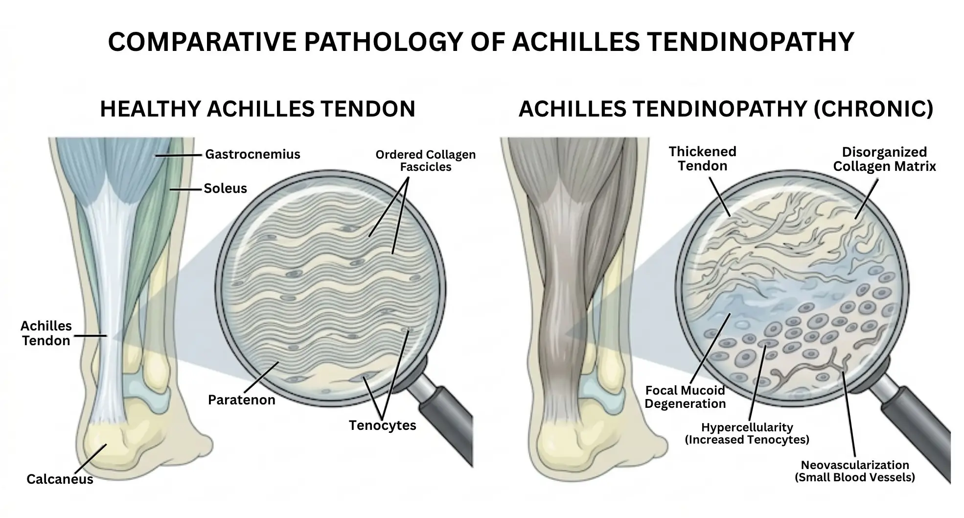
A tendon is a dense band of connective tissue that connects muscle to bone and transmits force from one to the other during every movement you make. Tendons look simple — whitish, fibrous, rope-like — but their internal architecture is precisely organized. The dominant structural protein is type I collagen, the same tough structural protein found in bone and skin, arranged in tightly packed parallel bundles. That organization is what gives tendons their extraordinary tensile strength: the Achilles tendon alone can withstand loads of several times body weight during running.
Woven into those collagen bundles are specialized cells called tenocytes (ten-oh-sites), whose main job is to maintain the collagen framework, sense mechanical load, and initiate local repair when small amounts of damage occur. Under normal circumstances, this works well enough. Tendons tolerate years of repetitive loading without permanent structural change.
Where Tendinopathy Begins — and Why It Doesn’t Resolve
When load exceeds the tendon’s capacity to repair — whether from sudden overload, accumulated microtrauma, age-related decline in cellular repair capacity, or altered movement mechanics — the collagen structure begins to break down. In healthy repair, type I collagen (strong, organized) is replaced in kind. In tendinopathy, the repair process is disrupted: type III collagen (weaker, less organized) is substituted instead. The ordered parallel structure of the fibers is lost, replaced by a disorganized, tangled matrix.
Critically, this is not a simple inflammatory injury. Biopsy studies of chronic tendinopathy consistently find reduced or absent inflammation at the cellular level — there are relatively few immune cells present. What they do find is structural disorganization, increased ground substance (the gel-like material between collagen fibers), abnormal cell populations, and the ingrowth of new blood vessels and nerve endings into regions that should have neither. This ingrowth of nerves is now thought to be one of the primary sources of the chronic pain that accompanies tendinopathy.
The central clinical problem is that this structural deterioration is largely self-sustaining. Tendons have an extremely limited blood supply — and unlike bone or muscle, they cannot easily call on the body’s standard repair systems. Tenocyte populations decline with age. In a chronically degenerated tendon, the remaining tenocytes have reduced capacity to produce normal collagen. The body produces scar-like tissue in its place, which weakens the tendon further and perpetuates both the structural problem and the pain.
What Conventional Tendinopathy Treatments Can and Cannot Do
Physiotherapy, Injections, and Shockwave: What Works and What Doesn’t
The current first-line treatment for tendinopathy is exercise rehabilitation — specifically a structured program of progressive tendon loading, which has the best available evidence for long-term outcomes. This is not dismissible: appropriate loading can stimulate collagen remodeling, reduce pain, and restore function, particularly in early-to-moderate tendinopathy. It should be the starting point, and often the majority, of any treatment plan.
Beyond exercise, the options narrow. Cortisone (corticosteroid) injections are widely used for pain relief. They can reduce swelling and discomfort in the short term, but a consistent finding in research is that their benefits often fade within weeks to months, and repeated injections may actually impair tendon integrity over time by inhibiting the cells responsible for collagen production. Platelet-rich plasma (PRP) — a concentration of platelets derived from the patient’s own blood, injected into the tendon — has attracted considerable attention as a more biologically active option. The evidence for PRP in tendinopathy, however, remains mixed: some controlled trials show meaningful improvement over placebo; others do not. The results appear to depend heavily on the specific tendon being treated, the stage of degeneration, and the preparation technique used.
Extracorporeal shockwave therapy (ESWT) — the application of acoustic pressure waves to the affected tendon — has reasonable evidence for certain tendinopathies, particularly calcific shoulder tendinopathy and mid-portion Achilles tendinopathy. It does not work for everyone, and its benefits are thought to be primarily through disrupting pathological nerve growth and stimulating a controlled local inflammatory response rather than through tissue regeneration.
The Surgical Ceiling for Tendinopathy
When conservative measures fail — and for a meaningful proportion of patients they do, with up to 45% of Achilles tendinopathy patients ultimately considering surgery — tendon debridement or repair is offered. Surgery removes the degenerated tissue and, in some cases, may stimulate a fresh repair response. The evidence for surgical outcomes in chronic tendinopathy is modest, recovery is prolonged, and the procedure itself carries risks including infection, nerve injury, and the risk that the repaired tendon develops further degeneration over time.
The practical gap that stem cell therapy is attempting to occupy is the same one described across musculoskeletal conditions: patients who have moved past the point where physiotherapy and injections provide durable relief, but who are not ready or not suited for surgery — and for whom no current treatment directly addresses the structural breakdown at the tissue level.
Clinical Data: What Studies Actually Show About Stem Cells and Tendinopathy
Key Published Trials and Their Numbers
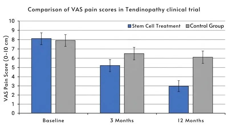
The evidence base for stem cell therapy in tendinopathy is in earlier stages than for conditions such as osteoarthritis or heart failure. There are no large Phase III randomized controlled trials yet. What exists is a body of Phase I and Phase II data, alongside well-designed smaller controlled trials, that provides meaningful — though not yet definitive — signal
A prospective, double-blind randomized controlled trial by Rodas and colleagues enrolled 20 patients with patellar tendinopathy that had not resolved after at least four months of conservative treatment. Ten patients received a single ultrasound-guided injection of autologous bone marrow-derived MSCs; ten received PRP as an active comparator. At the primary follow-up point, the MSC group demonstrated greater structural recovery on tendon ultrasound imaging than the PRP group — a meaningful finding because it suggests a structural, not merely symptomatic, effect.
A prospective case series by Pascual-Garrido and colleagues followed eight patients with chronic patellar tendinopathy — all of whom had failed at least six months of conservative treatment — who received ultrasound-guided injections of autologous bone marrow mononuclear cells. At five-year follow-up, significant improvement in the International Knee Documentation Committee (IKDC) functional score was documented in all patients. The five-year timeframe is notable: it suggests the benefit may be durable rather than simply a short-term response.
For the Achilles tendon, a 2024 first-in-human safety study published in Scientific Reports (Goldberg et al.) enrolled ten patients with mid-portion Achilles tendinopathy and injected autologous bone marrow-derived MSCs directly into the tendon. At 24-week follow-up, no serious adverse events were recorded, and functional outcome scores showed improvement from baseline across the group.
For rotator cuff tendinopathy, a controlled trial by Kim and colleagues assigned 12 patients with partial rotator cuff tears to MSC injection combined with PRP versus physiotherapy alone. The MSC-treated group showed significant reduction in pain scores and meaningful recovery of shoulder mobility at the three-month assessment compared to the physiotherapy-only group.
Regarding allogeneic cell sources — the approach used in Malaysia — a small uncontrolled trial of 12 patients with chronic lateral epicondylosis (tennis elbow) who received injections of allogeneic adipose-derived MSCs reported that treatment was safe and produced improvements in elbow pain, grip performance, and structural imaging at one year. This is an important data point because it demonstrates that allogeneic (donor-derived) cells can be used in tendon tissue without triggering rejection, expanding the practical options for patients.
What the Numbers Mean and What They Don’t
Taking this evidence together: MSC injections into degenerated tendons appear, in the available trials, to produce pain reduction and structural improvements that are more pronounced and more durable than control conditions in several tendon locations. The Pascual-Garrido five-year follow-up data is particularly encouraging as an indicator of durability.
However, the sample sizes in most trials are small. Very few trials include a proper placebo control, which matters because tendon pain can fluctuate over time regardless of treatment. The available evidence is Level 3 on the clinical evidence hierarchy — meaningful, but not yet strong enough to make a definitive recommendation in favor of or against this approach for all comers. We believe patients deserve to know this before committing to treatment, and not after.
Three Mechanisms: How MSCs Address Tendon Degeneration
Mechanism 1 — Calming the Disorganized Repair Environment
Mesenchymal stem cells (MSCs) — the cell type used in our treatment program — are not a form of pain reliever. They do not block a receptor or reduce a chemical signal the way cortisone does. Instead, they act as biological coordinators: releasing signaling molecules that shift the local tissue environment toward repair rather than continued degeneration.
In tendinopathy, one of the key problems is that the immune cells present in the tendon — particularly macrophages, which are the body’s general-purpose cleanup and repair coordinators — become stuck in a destructive “inflammatory” mode. MSCs secrete cytokines (chemical messengers between cells) that convert these macrophages from their destructive state to a tissue-remodeling state. This shift from destructive to constructive macrophage behavior is thought to be one of the primary reasons why patients treated with MSC injections report qualitatively different and more sustained relief than they experienced from steroid injections — the underlying cellular environment is being modulated, not temporarily suppressed.
Mechanism 2 — Supporting Tendon Cells and Collagen Production
The degenerated tendon lacks enough healthy tenocytes to produce the type I collagen needed for structural recovery. MSCs address this at two levels.
First, they secrete growth factors — proteins that stimulate the survival and activity of the tenocytes still present in the tendon. Think of this as sending reinforcements to support a depleted construction crew, rather than replacing them entirely. Second, research has shown that MSCs can themselves begin to adopt tendon-like characteristics in the right biochemical environment, contributing directly to collagen production alongside the existing tenocyte population.
The clinical consequence of this dual action, in patients who respond, can be detectable improvement in tendon structure on ultrasound or MRI — thicker collagen signal, more organized fiber alignment, and reduced hypoechoic regions (the dark patches on ultrasound that indicate degenerated tissue).
Mechanism 3 — Addressing Pathological Nerve and Blood Vessel Ingrowth
One of the least discussed — but clinically important — features of chronic tendinopathy is the abnormal ingrowth of new blood vessels and accompanying nerve fibers into regions of the tendon that should have neither. These nerve endings are thought to be a major source of the deep, persistent aching that characterizes chronic tendinopathy, particularly the pain that occurs at rest or during the night.
MSCs release molecules that inhibit the abnormal signaling driving this vascular and nerve ingrowth — a process called pathological angiogenesis. By reducing the signals that recruit abnormal vessels and nerves, MSCs may interrupt one of the primary pain-generating mechanisms in chronic tendinopathy, rather than simply blocking pain signals downstream.
Autologous vs. Allogeneic: Cell Source Matters in Tendinopathy Treatment
The Autologous Approach: Using the Patient’s Own Cells
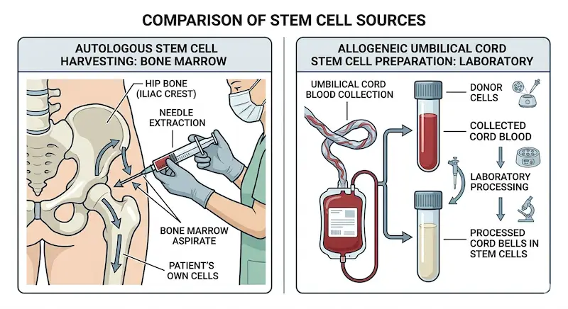
Most stem cell clinical trials in tendinopathy, and most clinics currently offering this treatment in Japan, use autologous MSCs — cells harvested from the patient’s own body, typically from bone marrow (drawn from the hip bone) or adipose (fat) tissue, then processed and injected back.
The principle advantage is clear: because the cells come from the patient themselves, immune rejection is not a concern. No matching or immunosuppression is required.
The limitation is equally clear. A 45-year-old with a two-year history of Achilles tendinopathy, chronic degeneration, and possibly other musculoskeletal conditions does not have the same stem cells they had at 20. Research consistently shows that MSCs from older donors — and those from individuals with chronic musculoskeletal disease — have reduced proliferative capacity, produce fewer beneficial growth factors, and have weaker immunomodulatory activity. The cells most needed for tendon repair may be the least available from the patient’s own aging tissues. Additionally, the harvesting procedure itself (bone marrow aspiration or liposuction) is invasive and adds a procedural step before any tendon treatment begins.
The Allogeneic Cord-Derived Approach Used in Malaysia
We use allogeneic MSCs derived from umbilical cord tissue — specifically the Wharton’s jelly layer surrounding the cord — donated by healthy mothers at the time of routine delivery. These cells are among the youngest, most potent MSCs available. They have not been exposed to years of repetitive mechanical load, inflammatory signaling, or pharmaceutical interventions. Their proliferative capacity and immunomodulatory output are measurably higher than cells from adult donors.
Crucially, umbilical cord MSCs express low levels of the surface proteins that normally trigger immune rejection. In every published clinical trial using allogeneic MSCs — including the lateral epicondylosis trial involving 12 patients — no immune rejection has been observed. The clinical safety of these cells in musculoskeletal tissue has been established across multiple studies.
From a practical standpoint, no harvesting procedure is required. The cells arrive in standardized, quality-tested preparations. Patients do not undergo an additional invasive step before treatment begins, and the cell dose administered can be verified for potency before injection.
The evidence does not yet firmly establish that allogeneic cord-derived MSCs are superior to autologous cells for tendinopathy specifically — the trial data comparing the two directly in this condition does not yet exist at scale. What the evidence does support is that allogeneic cord-derived cells are safe, biologically active, and offer a practical advantage for older patients and those whose own cells may be functionally compromised by age or prior disease.
Who Is — and Is Not — a Suitable Candidate for Tendinopathy Stem Cell Therapy
Profiles That Tend to Respond Well
Tendinopathy stem cell therapy shows the most promise in patients who share several characteristics. The underlying diagnosis is confirmed structural tendinopathy — typically by ultrasound or MRI showing focal degeneration, disorganized collagen signal, or hypoechoic (abnormal dark) regions within the tendon body. The patient has completed a meaningful period of structured physiotherapy (typically at least three to six months) without achieving durable resolution. At least one injection-based treatment — whether cortisone, PRP, or both — has been tried and has either failed to provide meaningful improvement or provided only short-term relief.
The tendon is degenerated but not fully ruptured. There is still structural continuity and tendon tissue remaining for biological repair processes to work with. Active inflammation is not the dominant finding on imaging — the characteristic picture is degenerative change rather than acute inflammatory tendinitis. And the patient’s overall health does not include active malignancy, uncontrolled autoimmune disease, or systemic infection.
In terms of the tendons themselves, the available clinical data is most directly relevant to mid-portion Achilles tendinopathy, patellar tendinopathy (jumper’s knee), rotator cuff tendinopathy, and lateral epicondylosis (tennis elbow). We do not currently have strong clinical trial data for all tendon locations.
Cases Where Stem Cell Therapy Is Unlikely to Help
A complete tendon rupture — not degeneration, but full structural failure — is a different clinical situation and is generally best addressed surgically. Stem cell therapy cannot bridge a gap between tendon ends or substitute for the mechanical repair a rupture requires.
Tendinopathy that is part of a widespread inflammatory arthritis — such as rheumatoid arthritis-associated tendinopathy — requires treatment of the underlying systemic condition alongside any local tendon intervention. Our rheumatoid arthritis stem cell program addresses the systemic inflammation that drives this pattern.
Patients with insertional tendinopathy (at the point where the tendon meets the bone) tend to respond less predictably than those with mid-substance degeneration, because the biology at the bone-tendon junction involves additional structural complexity.
Patients who need rapid functional recovery — for example, professional athletes with competition obligations within weeks — may find the gradual timeline of stem cell-mediated repair poorly matched to their situation. This is an honest limitation we raise in every consultation.
One Patient’s Recovery: A Tendinopathy Case From Our Malaysia Clinic
Background and Decision to Pursue Treatment
A woman in her early fifties, an avid recreational runner who had been managing bilateral Achilles tendinopathy for three years. She had completed two structured physiotherapy programs, received four cortisone injections across both tendons over two years (with diminishing relief from each), and one round of PRP. At the point she contacted us, her MRI showed focal mid-substance degeneration with intratendinous signal change at both Achilles tendons. Her pain at its worst was a 7 out of 10 on the standard pain scale, and she had reduced her weekly running mileage by approximately 80%. Her orthopedic surgeon had discussed surgical debridement as the next option.
After reviewing her imaging and treatment history remotely, we confirmed she was a realistic candidate. The tendons showed structural degeneration but remained intact. She had genuinely exhausted conservative options. We were clear before she traveled that we could not predict her individual response, and that a meaningful proportion of patients in her clinical situation do not achieve significant improvement.
What Changed — and What Didn’t
The first six weeks produced no noticeable change. This is expected and we had communicated it in advance.
By week eight, she reported that the first steps out of bed in the morning — historically her worst pain moment — had become noticeably less severe. By the three-month mark, her self-reported pain at walking speed was a 3 out of 10, and she had begun returning to light jogging (20-minute sessions). At six months, follow-up ultrasound showed improvement in the echogenicity (the texture and organization visible on ultrasound) of the degenerated regions in both tendons, with less prominent hypoechoic change compared to the pre-treatment baseline. Her functional outcome scores had improved substantially.
She has not returned to her prior training volume without discomfort — her tendons will require continued careful loading management and physiotherapy. Surgery has not been required or discussed further. The outcome she describes is not a return to a pain-free tendon, but a meaningful reduction in daily pain and a restoration of the ability to run at a recreational level — which was the primary goal she brought to us.
This is an anonymized account based on actual clinical experience. Individual results vary and cannot be guaranteed.
What to Realistically Expect in the Recovery Period
We communicate this clearly before every treatment: the first four to eight weeks will likely produce little or no noticeable change. Stem cell therapy works through a slower biological process than cortisone or PRP — the cells need time to engage with the local environment, modulate the inflammatory and cellular balance, and begin supporting collagen repair. Most patients who respond begin to notice change between weeks six and fourteen.
Early improvement typically comes in the form of reduced morning stiffness, less pain at low levels of activity, and a smaller “spike” of pain after activity. Structural changes on imaging, where they occur, usually become visible by the six-month scan. Patients who have not noticed any meaningful change by the six-month mark have generally not gone on to show changes at twelve months — which is honest information for anyone setting expectations at the outset.
Honest Risks and Limitations of Tendinopathy Stem Cell Therapy
Known Side Effects and Safety Data
The safety profile of MSC injection into tendon tissue is generally reassuring across the published literature. In the Goldberg 2024 Achilles tendinopathy safety study, no serious adverse reactions were recorded across ten patients at 24-week follow-up. In the allogeneic lateral epicondylosis trial, no immune reactions were observed in any of the 12 participants over one year.
The most commonly reported effects following tendon MSC injection are temporary and localized: injection-site pain, mild swelling, and short-term increased tendon discomfort in the first one to two weeks. These typically resolve without intervention. Because allogeneic MSCs express low levels of the surface proteins that trigger immune rejection, serious allergic or rejection responses are rare — but they are a theoretical risk with any biological therapy, and patients should be monitored accordingly.
Infection at the injection site is a risk inherent to any tendon injection, including routine cortisone and PRP procedures. We perform all injections under ultrasound guidance with strict sterile technique, which minimizes but does not eliminate this possibility.
What Stem Cell Therapy Cannot Do for Tendinopathy
There are clear limits to what this treatment can achieve, and we want to name them directly.
Stem cells cannot bridge a complete tendon rupture. If the tendon has fully torn — whether recently or through progressive degeneration leading to complete structural failure — biological injection therapy is not the appropriate first intervention. Surgical repair addresses the mechanical problem that biology cannot.
Not every patient responds. Based on published trial data and our clinical experience, a meaningful proportion — estimated at 20 to 35% across tendon types — do not achieve clinically significant improvement. We cannot predict with certainty who will respond. Pre-treatment imaging, the degree of existing degeneration, patient age, and biological factors we cannot yet fully characterize all influence outcomes.
The improvements documented in available trials are real but not complete. Patients should expect reduced pain and improved function — not a restored-to-new tendon. Continued physiotherapy and load management remain important after treatment, not optional additions.
Long-term data beyond three years is limited. The Pascual-Garrido patellar tendinopathy cohort provides a five-year observation, but this is one of very few such datasets. For most tendon locations, we do not yet have robust data on whether improvement at twelve months persists at five years, or whether re-treatment becomes necessary.
Anyone presenting this treatment as a guaranteed solution is not giving you an accurate picture.
FAQ About Tendinopathy Stem Cell Therapy
Cortisone reduces inflammation quickly — typically producing noticeable relief within days — but it does not repair the damaged tendon tissue. Its effects are often temporary, and repeated injections may impair the tendon’s structural integrity over time. MSC therapy works more slowly, over weeks to months, and aims to support actual biological repair of the tendon rather than suppressing symptoms.
Most patients at our clinic receive a single injection session per affected tendon. Depending on the number of tendons involved and the degree of degeneration, additional sessions may be discussed, but the default is a single focused treatment followed by structured follow-up.
Yes — and we actively encourage it. MSC therapy is not a replacement for structured tendon rehabilitation; it is a biological support for the repair processes that physiotherapy also promotes. In our follow-up program, patients work with a physiotherapist to progressively reload the tendon as recovery allows.
No. Stem cell therapy for tendinopathy in Malaysia is a self-funded treatment. We recommend patients confirm the absence of insurance coverage before proceeding and factor the full cost into their planning.
We advise against forming a conclusion before the six-month mark. The biological timeline of MSC-mediated tendon repair means that early absence of change is not a reliable indicator of eventual outcome. Patients who report no change at six months, however, have generally not gone on to show meaningful improvement at twelve months, and this is discussed honestly at that follow-up point.
What Comes Next
If you have reached this point, you are likely someone who has genuinely worked through the standard treatment options and is asking a serious question about whether this path makes sense for your specific situation.
We offer free online consultations where we review your imaging and treatment history and give you an honest assessment of whether your case falls within the range where published data suggests benefit is realistic. Many people use that conversation simply to understand their options more clearly before making any decision. If we do not think you are a realistic candidate, we will say so in that first call.
References
- Mao XF, Zhang XQ, Mao HJ, Yao ZY. “Advances in mesenchymal stem cells therapy for tendinopathies.” Journal of Orthopaedic Translation. 2024;44:30–40. https://doi.org/10.1016/j.jot.2023.11.002
- Goldberg AJ, Masci L, O’Donnell P, et al. “Autologous bone marrow derived mesenchymal stem cells are safe for the treatment of Achilles tendinopathy.” Scientific Reports. 2024;14:11421. https://doi.org/10.1038/s41598-024-61399-3
- van den Boom NAC, Winters M, Haisma HJ, Moen MH. “Efficacy of stem cell therapy for tendon disorders: a systematic review.” Orthopaedic Journal of Sports Medicine. 2020;8(4):2325967120915857. https://doi.org/10.1177/2325967120915857
- Pascual-Garrido C, Rolon A, Makino A. “Treatment of chronic patellar tendinopathy with autologous bone marrow stem cells: a 5-year-followup.” Stem Cells International. 2012;2012:953510. https://doi.org/10.1155/2012/953510
- Quintero D, Perucca Orfei C, Kaplan LD, de Girolamo L, Best TM, Kouroupis D. “The roles and therapeutic potential of mesenchymal stem/stromal cells and their extracellular vesicles in tendinopathies.” Frontiers in Bioengineering and Biotechnology. 2023;11:1040762. https://doi.org/10.3389/fbioe.2023.1040762
- Chen Z, Chen P, Zheng M, et al. “Challenges and perspectives of tendon-derived cell therapy for tendinopathy: from bench to bedside.” Stem Cell Research & Therapy. 2022;13:345. https://doi.org/10.1186/s13287-022-03113-6
- Wang JH. “Mechanobiology of tendon.” Journal of Biomechanics. 2006;39(9):1563–1582. https://doi.org/10.1016/j.jbiomech.2005.05.011
- Docheva D, Müller SA, Majewski M, Evans CH. “Biologics for tendon repair.” Advanced Drug Delivery Reviews. 2015;84:222–239. https://doi.org/10.1016/j.addr.2014.11.015